High quality ultrasound provides essential information enabling early detection of abnormalities and ensures timely medical intervention.
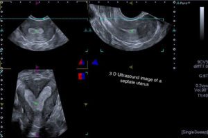
A pelvic ultrasound is a non-invasive diagnostic imaging procedure which is used to assess and visualize the female pelvis. Ultrasound produces detailed images of the uterus, endometrium, cervix, bladder, ovaries and adjacent pelvic structures.
A transvaginal ultrasound scan is the technique for quick, effective assessment and diagnosis of various conditions. It is the initial method favoured by clinicians as it is completely safe, posing minimal risks to the patient.
A gynaecological ultrasound scan is requested when abnormalities of the female reproductive system are suspected on the basis of certain symptoms that are concerning you and/or your doctor. The scan may provide you with quick answers and peace of mind.
Transvaginal ultrasound is a screening test for endometrial cancer. It is not a screening test for cervical cancer nor is it a screening test for ovarian cancer. However it can reassure you that your ovaries are normal. It is also part of the investigation of infertility and monitoring of infertility treatment strategies.
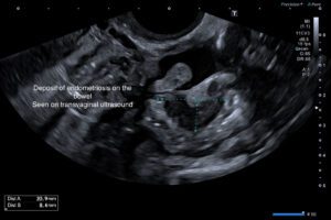
Transvaginal ultrasound (TVUS) is a valuable tool for the diagnosis and staging of endometriosis. The 2022 update of the European Society of Human Reproduction and Embryology (ESHRE) guidelines for diagnosis and management of endometriosis suggests that laparoscopy should no longer be considered the gold standard in diagnosis, favouring imaging as the first-line investigation.
An endometriosis is a complex scan comprising of a consultation, the scan itself and a detailed consultant report.
In the early weeks of pregnancy, TV ultrasound is used to confirm that:
Early pregnancy scans are typically conducted between 5.5 and 11 weeks of gestation. The scan is an internal TV scan prior to 8 weeks.
The changes in the uterus in a normal early pregnancy occur in a very organised sequential manner:
The pregnancy sac which is called the gestational sac is the first structure seen and within a few days changes start to occur within that sac. The second structure seen on TV ultrasound is the yolk sac, which provides support for the pregnancy during the first 12 weeks. The embryo then appears adjacent to the yolk sac, it is often just 1.5 to 2mm when it is first seen and it grows at a rate of 1.2mm per day. The crown-rump length (CRL) is measured to estimate the embryo’s age.
If the scan is performed too early , that is before 6 weeks, the embryonic heartbeat may not be detectable, as it is often not visible at this stage.
Scans performed at 10 to 11 weeks generally provide visibility of the embryo’s head, limbs, hands, and feet.
The ovaries, uterus, and cervix are also examined as part of a comprehensive evaluation.
No preparation for the scan is necessary.
No GP referral is required.
Scanning in infertility encompasses a number of assessments
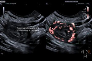
A follicle tracking scan measures the size of active ovarian follicles. Follicles are fluid-filled structures in the ovaries that may contain an egg, and their size can be used to estimate when ovulation will occur. A scan is performed to assess the timing of ovulation, providing information that can be used in both natural conception cycles and fertility treatments.
The ovaries are however not evaluated in isolation. The lining of the womb ie the endometrium is also assessed. An endometrial thickness scan measures the womb lining (endometrium) to track changes during a normal cycle, correlate ovulation with ovarian appearances, or monitor responses to fertility drugs used in treatment.
Follicle tracking and endometrial measurement scans are typically done three times per menstrual cycle. The first, a detailed gynaecological scan, occurs between days 3 and 5 of your period (with day one being the start of bleeding). Further scans, focusing on monitoring the natural changes of the ovaries and endometrium, (or treatment induced changes) follow at 1–2 day intervals as needed.
These scans therefore
A GP referral is not required and you will receive your results on the day of the scan and a detailed report to take away for your gynaecologist or fertility unit, the report can be emailed to you for your convenience (with your permission).
Transvaginal ultrasound (also known as an internal ultrasound, or vaginal ultrasound) is where images are obtained with a transducer or probe that is placed into the vaginal cavity. This is THE method of choice and MUST be the initial investigation. A full bladder is NOT required.
Transabdominal scan (through the abdomen). This is an external ultrasound scan, as the images of the pelvic organs are obtained through the abdomen using a full bladder. This is an additional secondary part of the investigation and will include the kidneys, which can be affected by pelvic abnormalities. A full bladder is not usually required.
Most women believe the internal scan is less uncomfortable than a cervical smear test or internal examination performed by your doctor or gynaecologist but you may ask to stop the scan at anytime.
The whole procedure is usually completed within 20 to 25 minutes. An endometriosis assessment takes longer, usually 30 to 40mins.
Eat and drink as you normally would. For most women no preparation is necessary (no full bladder needed) as the initial scan is an internal scan.
If a full bladder is necessary (under certain conditions an internal scan cannot be performed) you will be asked to drink 1.5 pints of water 90 minutes before the scan.
After an ultrasound scan examination there is no restriction on activities.
There are 2 forms of referral:
Medical practitioner referral: means that you have a referral letter from your doctor. A referral form is available which your doctor can complete and email to us at: hera.imaging@healthmail.ie
Your doctor may also post us your referral.
Self referral: referral form is available which you can complete and email to us at: office@heraimaging.ie
After the scan you will be informed of the results.
A written report is sent to you or your referring doctor within 2 working days.
We would be happy to assist you if you have any further questions.
For general questions or appointment booking, you can contact our office via email: office@heraimaging.ie or by phone: +353 85 711 2438
For questions or concerns relating to your scan, please contact Dr. Coady via email: annemarie@heraimaging.ie